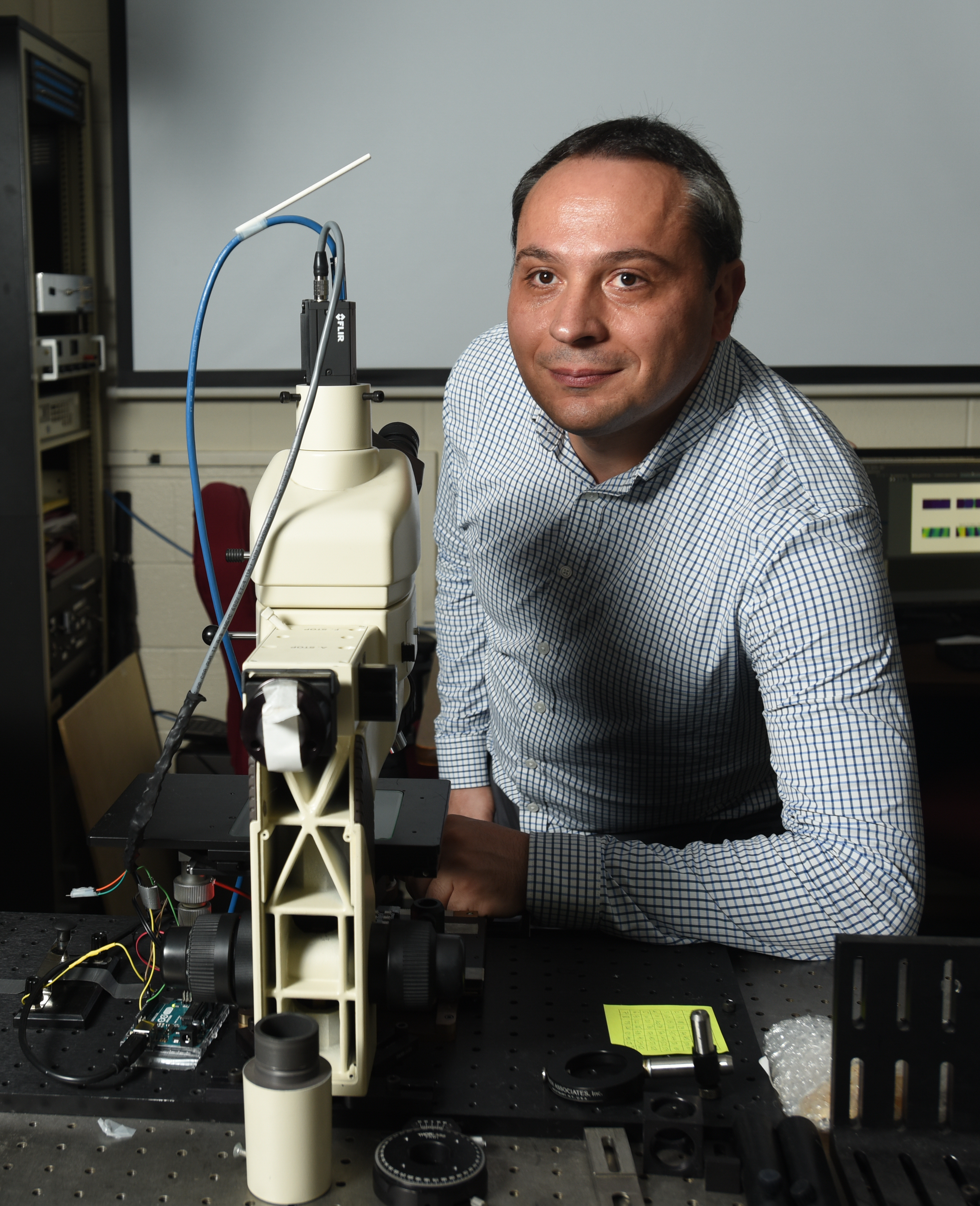
George Nehmetallah, Ph.D., Ordinary Professor in the Department of Electrical and Computer Engineering at The Catholic University of America, has been awarded a U.S. Patent for his groundbreaking invention: Fourier Ptychographic Microscope Systems and Methods. This innovation represents a major advancement in high-resolution imaging, combining computational microscopy with artificial intelligence to deliver enhanced diagnostic capabilities.
Traditionally, polarized light microscopy has been used to analyze birefringent specimens—biological materials like tissues and cells that change the direction of light depending on its orientation. This technique is widely employed in pathology for its ability to reveal structural details not visible through conventional imaging methods.
Fourier ptychographic microscopy (FPM), on the other hand, is a cutting-edge computational imaging technique that increases both resolution and field of view by capturing multiple images at varying illumination angles. However, traditional FPM treats light as a scalar field and does not incorporate valuable polarization information—an important limitation in biomedical imaging.
Nehmetallah’s invention overcomes this by integrating polarization sensitivity into the FPM framework. His team developed a novel method called Deep Learning-based Polarization Fourier Ptychographic Microscopy (DL-PFPM). Utilizing a Generative Adversarial Network (GAN) architecture, the technique reconstructs high-resolution, wide field-of-view images that include quantitative information on birefringence retardance and orientation—data that can be extracted from a single recovered complex field image.
“This approach enables detailed polarimetric analysis at a much higher resolution, using AI to uncover information that was previously inaccessible with conventional methods,” said Nehmetallah.
The potential applications of this technology are wide-ranging. In the semiconductor industry, it can be used to trace defects in crystalline components—essential in the production of computer chips and mobile devices. In clinical pathology, it could revolutionize early-stage disease detection. For instance, DL-PFPM allows clinicians to detect changes in collagen structures associated with early-stage cancers or parasitic infections without the need for special staining or sample preparation.
This innovation underscores Catholic University’s growing contributions to biomedical engineering and artificial intelligence, paving the way for enhanced diagnostic tools that are faster, more accurate, and less invasive.